The adrenal gland
Two small triangular glands, one on top of each kidney. The outer layer (cortex) and the inner core (medulla) make different hormones, which is why adrenal conditions can present in very different ways.
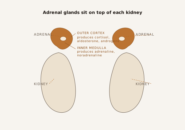
Where they sit
Each adrenal gland sits just above the kidney, tucked up under the diaphragm. The right gland usually sits close behind the liver and the inferior vena cava (the large vein returning blood to the heart). The left gland sits near the spleen, pancreas tail and left kidney vein. These close relationships matter when planning surgery, because safe access and vessel control are part of why the robotic approach is now widely used.1
Cortex - the outer layer
The cortex makes three groups of steroid hormones:
- Cortisol helps the body handle stress, keeps blood sugar steady, and has a role in blood pressure. Too much cortisol causes Cushing's syndrome.
- Aldosterone regulates blood pressure through sodium and potassium balance. Too much aldosterone causes Conn's syndrome (primary hyperaldosteronism).
- Adrenal androgens are weak sex hormones. Excess production is uncommon but can be a feature of some adrenocortical tumours.
Medulla - the inner core
The medulla makes catecholamines: adrenaline (epinephrine) and noradrenaline (norepinephrine). These drive the fight-or-flight response, tightening blood vessels and raising heart rate. A tumour of the medulla is called a phaeochromocytoma, and can release catecholamines in surges that cause dangerous blood pressure spikes.6
How hormone testing fits in
Because the two parts of the gland make different hormones, the workup for an adrenal mass routinely looks at all of them before an operation is planned:
- Plasma metanephrines (or 24-hour urinary metanephrines) for phaeochromocytoma.
- Aldosterone-to-renin ratio, especially in patients with high blood pressure or low potassium.
- Overnight 1 mg dexamethasone suppression test, or 24-hour urinary free cortisol, to screen for autonomous cortisol production.
These tests are usually organised by your GP or endocrinologist alongside imaging, and guide whether surgery is needed, how to prepare for it, and which specialist team should be involved.3
What removing one gland means
For most patients, the other adrenal gland takes over normal hormone production after surgery. If both glands are removed (uncommon, usually for bilateral disease or hereditary phaeochromocytoma), lifelong steroid replacement is required. Your team will discuss this in detail before the operation.